
Page 40 - Journal of Structural Heart Disease Volume 3, Issue 6
P. 40
Case Report 196
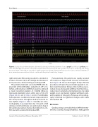
Figure 5. Cardiac invasive hemodynamics after mitral valve repair with measurement of aortic (purple) and left atrial (yellow) pres- sures. After MitraClip placement, overall improved left atrial pressures (mean 20 mm Hg with V waves up to 33 mm Hg) and arterial pressures (mean 76 mm Hg) are seen with no signi cant pressure changes during ventricular paced or native conducted rhythms. Of note, minimal pressure variations did not correlate with the patient’s ventilatory changes.
right ventricular (RV) pacing resulted in a marked el- evation of V waves (up to 67 mmHg) and decrease in aortic pressures, demonstrating worsening of MR.
The patient underwent successful PMVR with de- ployment of three MitraClips along the A1-2/P1-2 in- terface, with reduction of MR from severe to mild and a mean transmitral gradient of 3 mmHg (three-di- mensional planimetry valve ori ce area of 3.6 cm2) (Figure 2B, 2C, and 4, Video 3 and 4). Post-MitraClip hemodynamics con rmed dramatic improvement in left atrial pressures throughout both paced and na- tive rhythms (Figure 5, Table 1). Transthoracic echo- cardiography on postoperative day 1 showed a mild decrease in left ventricular size and modest drop in left ventricular ejection fraction from 60–65% to 45– 50% but with sustained mild residual MR.
Postoperatively, the patient was rapidly weaned o vasopressor support with recovery of renal func- tion (Figure 1B). He was discharged 10 days after the procedure, with plans for pacemaker upgrade to car- diac resynchronization therapy. One month post-pro- cedure, he was doing well, with New York Heart Asso- ciation class II symptoms and maintenance of a stable weight on an oral diuretic regimen with brain natri- uretic peptide of 126 pg/mL. Follow-up echocardiog- raphy con rmed no signi cant changes, with stable mild residual MR.
Discussion
Cardiac pacing is an established and e ective treat- ment for a variety of bradyarrhythmias. Although the
Journal of Structural Heart Disease, December 2017
Volume 3, Issue 6:192-198
