
Page 38 - Journal of Structural Heart Disease Volume 3, Issue 6
P. 38
Case Report 194
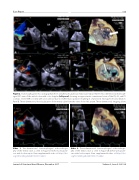
Figure 2. Transesophageal echocardiography before and after percutaneous mitral valve repair. Panel A. Two-dimensional midesoph- ageal 50° view of the mitral valve with color doppler (left panel) showing an approximate commissural view of the P3, A2, and P1 scallops. Severe MR is noted, with a broad base due to lea et malcoaptation resulting in a functional etiology from atrial dilation. Panel B. Three-dimensional live visualization of the mitral valve from the view of the left atrium. Three-dimensional imaging allows
Video 1. Two-dimensional transesophageal echocardiogra- Video 2. Three-dimensional transesophageal echocardiogra-
phy of the mitral valve as seen in Figure 2A before percutane- ous mitral valve repair. View supplemental video at https://doi. org/10.12945/j.jshd.2017.033.17.vid.01.
phy of the mitral valve as seen in Figure 2B before percutane- ous mitral valve repair. View supplemental video at https://doi. org/10.12945/j.jshd.2017.033.17.vid.02.
Journal of Structural Heart Disease, December 2017
Volume 3, Issue 6:192-198
