
Page 39 - Journal of Structural Heart Disease Volume 3, Issue 6
P. 39
195 Case Report
Figure 3. Invasive cardiac hemodynamics measuring simultaneous aortic (purple) and left atrial (yellow) waveforms before mitral valve repair. When the patient was in a ventricular paced rhythm, increased left atrial pressures with a mean of 30 mmHg with prom- inent V waves of up to 75 mmHg were noted. Simultaneous central aortic pressures decreased with ventricular pacing (mean 58 mmHg). Native ventricular conduction was associated with decreased left atrial pressures (mean 20 mmHg) with smaller V waves and enhanced arterial pressures (mean 68 mmHg). Ao = aortic pressures; LA = left atrial pressure.
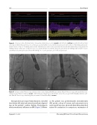
Figure 4. Intraprocedural uoroscopy, anteroposterior view, during MitraClip procedure. Panel A. Fluoroscopy performed after trans- septal puncture. Balloon dilation (circle) of the interatrial septum was performed to allow for passage of the MitraClip delivery cath- eter. Panel B. Fluoroscopy visualizing the placement of three MitraClips (arrows).
Intraoperative pre-repair hemodynamics revealed two distinct left atrial and arterial waveforms depend- ing on the patient’s cardiac rhythm, illustrating its electromechanical in uence on MR (Figure 3). Where-
as the patient was predominantly atrioventricular (AV) paced, a drop in V waves and concurrent rise in aortic waveforms was observed when the patient had native AV conduction. In comparison, resumption of
Nguyen H. L. et al.
Worsening MR from V-Paced Septal Dyssynchrony
