
Page 37 - Journal of Structural Heart Disease Volume 3, Issue 6
P. 37
193 Case Report
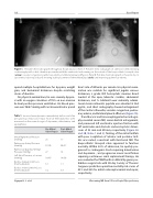
Figure 1. Portable chest radiographs throughout hospitalization. Panel A. Portable chest radiograph on admission demonstrating cardiomegaly with a dual-chamber pacemaker with the ventricular lead running on the outside of the bioprosthetic tricuspid valve (arrow), vascular congestion, pulmonary edema, and bilateral pleural e usions. Panel B. Portable chest radiograph on hospital day 12 (post-mitral valve repair day 6) showing stable placement of three MitraClips (circle) and improving pulmonary edema.
quired multiple hospitalizations for dyspnea, weight gain, and abdominal distension despite escalating doses of diuretics.
On physical examination, he was severely dyspne- ic with an oxygen saturation of 95% on non-invasive bi-level positive pressure ventilation. His blood pres- sure was 96/67 mmHg with an atrioventricular paced
Table 1. Invasive hemodynamics immediately before and after percutaneous mitral valve repair. Note all hemodynamics were measured on the same dosages of dopamine, dobutamine, and epinephrine.
Mean Right Atrial Pressure
(mmHg) 23 17
Pulmonary Artery Pressure
(mmHg) 67/30 60/27
Pulmonary Capillary Wedge
Pressure (mmHg) 27 18
Left Atrial Pressure, a/v waves
(mmHg) 18/65 14/30
Cardiac Output (L/min; via
thermodilution) 6.1 7.1
Cardiac Index (L/min/m2; via
thermodilution) 3.2 3.8
heart rate of 80 beats per minute. His physical exam- ination was notable for signi cant jugular venous distension, a grade II/VI holosystolic murmur heard loudest at the apex, bibasilar crackles, abdominal distension, and 1+ bilateral lower extremity edema. Serum brain natriuretic peptide was elevated at 364 pg/mL, and chest radiography showed enlargement of the cardiac silhouette, vascular congestion, pulmo- nary edema, and bilateral pleural e usions (Figure 1A).
Transthoracic and transesophageal echocardiogra- phy revealed severe MR, severe biatrial enlargement, and preserved left ventricular ejection fraction with left ventricular end-diastolic and end-systolic dimen- sions of 52 mm and 40 mm, respectively (Figure 2A and 2B, Video 1 and 2). Tenting of the mitral lea ets with poor coaptation of anterior and posterior leaf- lets was noted, consistent with functional MR. The bioprosthetic tricuspid valve appeared to function normally. Within 24 h of admission, he quickly pro- gressed to cardiogenic shock requiring dobutamine and dopamine , and progressive anuric renal failure requiring continuous renal replacement therapy. He was evaluated for PMVR with via MitraClip given pro- hibitive surgical risk with 30-day Society of Thoracic Surgeons predictive operative mortality risk scores of 43% and 36% for mitral valve replacement and repair, respectively.
Pre-Mitral Valve Repair
Post-Mitral Valve Repair
Nguyen H. L. et al.
Worsening MR from V-Paced Septal Dyssynchrony
