
Page 15 - Journal of Structural Heart Disease Volume 3, Issue 6
P. 15
171 Original Research Article
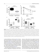
Figure 5. Comparison of vessel lumen diameter change, endothelialization, and vessel wall injury after intentional stent fracture between pre-stent and single stent groups. Panel A. Histomorphometric comparison of stented vessel lumen area 2 months after intentional stent fracture. The area of the stented vessel segments was signi cantly larger in the pre-stent group than in the single stent group. Panel B. The pre-stent group maintained larger angiographic lumen diameter, whereas the single stent group exhibited signi cant diameter loss due to stent in-folding and collapse 2 months after balloon angioplasty. Panel C. Pre-stenting allowed almost complete endothelialization 2 months after stent implantation, whereas endothelial coverage was signi cantly less in the single stent group due to luminal stent strut protrusion. Panel D. Comparison of vessel injury between pre-stent and single stent groups at short-term and mid-term evaluation. Vessel wall injury was similar at the time of stent fracture regardless of pre-stenting or unzipping without pre-stenting; however, injury score signi cantly improved 2 months after stent fracture in the pre-stent group but not in the single stent group. Data are shown as median, interquartile range, and range.
compared with the pre-stent group (Table 2). The de- gree of vessel diameter loss was subtle on traditional angiography (Figure 3A), but high-resolution radiog- raphy and rotational CT showed compromised vessel patency (Figure 3B and 33C) and revealed the mecha- nisms of vessel diameter loss in the single stent group as including stent in-folding, buckling, and collapse. This stent in-folding, buckling, and collapse was not present in the pre-stent group (Figure 4).
Consistent with the maintained vessel diameter as shown by angiography, radiography, and CT, the lu-
minal area of the stented vessel segments measured by histomorphometry was signi cantly larger in the pre-stent group (109 mm2 (89–141)) than in the single stent group (57 mm2 (47–73), P = 0.019; Figure 5A). The angiographic vessel diameter of stented vessel segments was similar to that of adjacent naïve ves- sel segments in the pre-stent group, whereas the single stent group demonstrated signi cantly larger luminal diameter loss. Two months after angioplasty, compared with the balloon diameter used for dilation (we only compared segments after dilation with 16
Bratincsák A. et al.
Pre-Stenting with Intentional Stent Fracture
