
Page 13 - Journal of Structural Heart Disease Volume 3, Issue 6
P. 13
169 Original Research Article
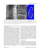
Figure 3. Reduced vessel patency and stent integrity after intentional fracture without pre-stenting. Panel A. Angiogram showing a small decrease in the diameter of the stented vessel segment compared with the adjacent area 2 months after intentional fracture and re-dilation of a 6-mm Cook Formula 418 stent with an ultra-high-pressure 16-mm Atlas balloon. Black arrows indicate the edges of the Cook stent. Panel B. High-resolution radiograph of the same segment showing fractured circumferential struts. Panel C. Down- the-barrel view of a three-dimensional rendition CT image of the same stent showing severely compromised luminal diameter in the antero-posterior dimension with struts protruding into the vessel lumen.
DIGI-VIEW 400, and micro CT was performed using a Scanco VivaCT40 System.
Histopathology. Perfusion- xed, excised arteries were further immersion- xed in 10% neutral bu ered formalin. After dehydration though a serial gradient of alcohols and clearing in xylene, tissues were in l- trated with methyl methacrylate and polymerized in a water bath. Plastic embedded arteries, with stents in place, were cut on a diamond-encrusted saw blade into 30–50 μm sections. Proximal distal sections of stented artery (taken at 25% and 75% of the stent length) were stained with hematoxylin and eosin us- ing routine techniques. Proximal and distal reference sections were trimmed from the xed arteries, pro- cessed using routine para n histology techniques, and stained with hematoxylin and eosin. Morpho- metric measurements of each cross-section included vessel lumen area. Histopathological evaluation of stented vessels was performed by a board-certi ed veterinary pathologist. Cross-sections within each stent were evaluated for injury using an established semi-quantitative scoring system based on indus- try-standard consensus recommendations [15]. At
each strut within a cross-section, vessel wall injury was scored as follows: 0 for no change in internal elas- tic lamina, 1 for rupture of internal elastic lamina, 2 for injury to tunica media, or 3 for injury to external elastic lamina and extending into or through the tuni- ca adventitia. Each vessel had two cross-sections. The score for each cross-section was the sum of all injury scores for that cross-section divided by the number of total struts in the cross-section. The injury score for each group is the injury score for all cross-sections for the group divided by the total number of cross-sec- tions for the group. Endothelialization was deter- mined by the histological presence of endothelium over each strut within a cross-section. The degree of endothelialization is the percentage of endotheliali- zed struts in the vessel.
Statistical Analysis
Descriptive statistical analysis was used to compare variables between pre-stent and single stent groups; data are expressed as median and interquartile range. Mann-Whitney tests were used to compare vessel lu- men area, balloon size, degree of endothelialization,
Bratincsák A. et al.
Pre-Stenting with Intentional Stent Fracture
