
Page 14 - Journal of Structural Heart Disease Volume 3, Issue 6
P. 14
Original Research Article 170
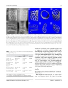
Figure 4. High-resolution radiographic and three-dimensional rendition CT images comparing pre-stenting at the time of stent fracture and single stent fracture. Intact stent integrity in the pre-stented segment maintained vessel patency, whereas loss of stent integrity after single stent fracture caused in-folding and compromised vessel lumen patency. Panels A, B, C, D, and E. Palmaz Genesis 1910 B stent implanted inside a Cook Formula 418 stent with simultaneous intentional fracture of the Cook stent. Images were taken 2 months after intentional fracture of the Cook stent. Images show intact internal stent integrity of the pre-stented vessel segment 2 months after implantation of the Palmaz Genesis stent with simultaneous intentional fracture of the 5-month previously implanted Cook stent. Panels F, G, H, I, and J. Cook Formula 418 stent 2 months after stent fracture (i.e., unzipping) without pre-stenting. Images show compromised stent integrity and vessel patency of the stented segment 2 months after intentional stent fracture and re-dila- tion of the 5-month previously implanted Cook stent.
Table 2. Comparison of outcome measures between the re-stent and single stent groups.
and vessel wall injury scores between groups; data are expressed as median and interquartile range. Mann-Whitney and Wilcoxon rank tests were used to compare the loss of luminal diameter from implanta- tion to mid-term between groups; data are expressed as median change (%) and interquartile range. Vessel wall injury scores were compared between groups using unpaired t-tests and Mann-Whitney post-hoc tests for nonparametric data. Analyses were per- formed using Excel (Microsoft, Redmond, WA), Graph- Pad Prism (GraphPad, La Jolla, CA), and Instat 3 (Graph Pad, San Diego, CA) software. P-values < 0.05 were considered statistically signi cant.
Results
Vessel Patency and Luminal Diameter after Intentional Stent Fracture
After intentional stent fracture, we found signif- icant vessel diameter loss in the single stent group
Re-stent group
Single stent group
p-value
Lumen area (mm2)* of stented blood vessels
Vessel diameter loss (%)*
2 months after dilation
Endothelialization (%) of stent struts
Vessel wall injury score per struts
108.9 (88.7- 140.7) (n=4)
44 (26-59) (n=4)
100 (89-100) (n=8)
0.3 (0.26-0.45) (n=8)
56.8 (47.3- 72.8) (n=4)
75 (61-85) (n=4)
73 (56-96) (n=8)
0.5 (0.35-0.63) (n=8)
0.0190 0.0065
0.0221
0.0673
Values expressed as median and interquartile range.
* Only the stents dilated with 16 mm balloon for diameter conformity
Journal of Structural Heart Disease, December 2017
Volume 3, Issue 6:165-175
