
Page 55 - Journal of Structural Heart Disease Volume 4, Issue 4
P. 55
157
Meeting Abstracts
systemic hypertension after 5±4 follow-up. Conclusions: Patients with aortic coarctation treated with stent at early age, can be successfully re-treated by balloon dilation or re-stenting after the completion their somatic growth.
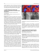
82. THREE-DIMENSIONAL PRINTING AND DYNAMIC TESTING PRIOR TO PULMONARY VALVE REPLACEMENT
R. Allen Ligon, Sassan Hashemi, Timothy Slesnick, Christopher Petit
Children's Healthcare of Atlanta - Emory University School of Medicine, Atlanta, USA
Background: Three-dimensional (3D) virtual and printed models have emerged as promising tools in the planning of interventions for patients with complex congenital heart disease (CHD). 3D replications can provide vital anatomic information to operators in planning pediatric cardiac inter- ventions both by optimizing the approach and possibly, deciding if a procedural approach is achievable. Typically, these 3D models are static representations of anatomy. We describe our approach with dynamic testing on 3D models of CHD prior to pulmonary valve intervention.
Methods: 9 patients underwent 3D modeling and printing prior to planned transcatheter pulmonary valve replace- ment (PVR) or surgical intervention. These patients under- went preprocedural cardiac magnetic resonance imaging (cMRI) or computed tomography (CT). From their imaging data, replications in the form of 3D virtual datasets (see gure) and 3D physical prints were generated. Dynamic evaluation of 3D models was performed with increasing diameter balloons to test for coronary artery (CA) compres- sion. Results of 3D modelling analysis were compared with actual procedural results during transcatheter PVR.
Results: Of the 9 patients who underwent 3D model- ing and printing, 4 were referred for surgical repair. The 5 remaining patients were or will be taken to the catheter- ization laboratory for transcatheter PVR. The dynamic 3D testing indicated CA compression in 3 patients and no CA compression in the remaining 2. In 2 of the patients with suspected CA compression, test dilation of the 3D ren- ditions at diameters of 16mm and 18mm were normal. However, CA compression was subsequently suggested during dilation with an increase to an 18mm or 20mm bal- loon, respectively (see gure). These patients were taken to the cath lab for PVR and during angioplasty of the con- duit were noted to have CA compression at the 18mm and 20mm balloon diameters, just as the 3D models had
predicted. These patients were ultimately referred for sur- gical PVR.
Conclusions: Anatomic 3D modelling is increasingly important in interventional and surgical planning for patients with CHD. However, dynamic 3D assessment may unveil limitations or potential complications, and should be considered an important adjunct to pre-procedural planning.
84. WHAT BALLOON SIZE SHOULD BE USED IN NEONATES WITH CRITICAL AORTIC STENOSIS?
Manish Malkar, Saadeh Jureidini
Saint Louis University, Saint Louis, USA
Background: The optimal size of the balloon for aortic val- vuloplasty (BAV) for critical congenital aortic valve stenosis (CCAS) has not been identi ed. An ideal balloon decreases the pressure gradient to safe level and minimizes aortic regurgitation (AR).
Methods: We retrospectively reviewed data from neo- nates with CCAS who underwent BAV from the year 1999 through 2018. We aimed to determine the balloon size that reduces pressure gradient across aortic valve to safe level and minimizes AR.
Results: Twenty consecutive neonates with CCAS under- went BAV in the rst 15 days of life. The balloon size used were ≤ 6 mm (group 6; n=12) and ≥ 7 mm (group 7; n=8). The birth weights were similar in both groups (P=0.1). Although the aortic valve annulus size in group 7 was
Hijazi, Z
21st Annual PICS/AICS Meeting
