
Page 53 - Journal of Structural Heart Disease Volume 4, Issue 4
P. 53
155
Meeting Abstracts
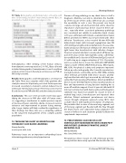
77. Table. Demographics and hemodynamics at baseline, with nitric oxide testing and after Fontan stent placement. Data are presented as median (intraquartile range) or n (%).
hemodynamics. After stenting of the Fontan stenosis, hemodynamics were repeated on 21% FiO2. Data collected included demographics, hemodynamics in each condition and percent increase in CI and percent decrease in PVR on iNO and post-stent.
Results: Demographics and hemodynamics are shown in the Table. There was complete relief of the gradient after stent placement in all patients; post-stent Fontan pressure increased secondary to transient contrast-induced rise in ventricular end-diastolic pressure. There was an increase in CI and decrease in PVR with iNO and after stent placement.
Conclusions: This case series provides novel data quan- tifying the decrease in PVR after relief of Fontan steno- sis, suggesting a mechanism to explain previous reports of decreased lower extremity edema, increased exercise tolerance and improvement in protein losing enterop- athy. Larger studies will likely con rm the signi cance of these ndings, but these data are a compelling addition to the long term management of this complex patient population.
78. TACKLING THE SHUNT LESION WITH LUNG PATHOLOGY: CASE BASED LEARNINGS
Neeraj Awasthy
max hospital, delhi, India
Pulmonary issues are an important confounding factors determining pulmonary artery hypertension
Presence of lung pathology in any shunt lesion is itself a diagnostic dilemma, not only to determine the feasilbil- ity of the closure of the cardiac defect but also ascertain the operability in such a case. We present case based scenarios (in large asd and nonresticted vsd) to show the importance of percutaneous device closure in these sub- sets , especially when associated pulmonary pathology was considered not suitable to undertake shunt closure .A 56 year old female with Ostium secundum Atrial Septal Defect presented in NYHA class IV presented with chest infection. Transthoracic echo revealed 30 mm ASD with bidirectional shunt, with tricuspid regurgitation max PG of 85 mmHg and right ventricle dysfunction. She was intu- bated and put on mechanical ventilator. CT chest showed right lower lobe bronchiectasis with infective changes. However repeated attempts to wean patient o bipap sup- port were unsuccessful. Subsequently cardiac catheteriza- tion and Balloon Occlussion of ASD revealed Qp/Qs ratio of 5:1 with step up in oxygen saturation of 19%. She under- went successful device closure for ASD with AMPLATZER septal occluder (36mm) (AGA Medical Corp., Minneapolis, MN, USA). Her physical activity and symptoms improved. Follow-up echocardiography after 5 months revealed nor- mal right ventricle contractility and no PAH. While initially chest infection prevented ASD device closure, underly- ing bronchiectasis which got worsened by asd shunt, pre- vented her from weaning o ventilator. Atrial Septal Defect device closure thereby helped to reduce pulmonary blood ow and improvement in lung function enabling her to wean o ventilator support. Case 2: 8 year old child with 10 mm non restricted vsd with dextrocardia and Kartageners syndrome was found not suitable for closure in view of marked lung issues, bronchospasm and repeated chest infection. Patient underwent percutaneous vsd device closure through antegrade approach using 10/12 duct occluder device with signi cant resolution of symptoms. We postulate that in view of decease lung segment, rest of the lung segment get ooded with the ow through the shunt lesions and compromise lung compliance. The closure of defects in such situations greatly facilitates the symptomatic improvement.
79. PERCUTANEOUS CLOSURE OF A LEFT VENTRICULAR PSEUDOANEURYSM IN A REDO POST MVR PATIENT: LEARNINGS FROM THE LARGEST REPORTED CASE
Neeraj Awasthy
Max Saket, Delhi, India
Percutaneous device closure of a left ventricular pseudo- aneurysm have been rarely reported. We describe the case
Hijazi, Z
21st Annual PICS/AICS Meeting
