
Page 26 - Journal of Structural Heart Disease Volume 3, Issue 6
P. 26
Case Report 182
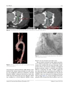
Figure 4. Three-dimensional TEE showing a heavily calci ed mitral valve with severe stenosis..
Figure 5. TEE four chamber view with color doppler showing severe mitral stenosis.
reconstruction software (Pixmeo SARL, Bernex, Swit- zerland). This analysis demonstrated an aortic annu- lus area of 480 mm2, which was suitable for a 26-mm Edwards SAPIEN 3 (Edwards Lifesciences, Irvine, CA, USA) valve. The mitral valve area was 286 mm2, which was suitable for a 29-mm Edwards SAPIEN 3 valve.
Figure 6. Fluoroscopy clip showing deployment of the SAPIEN 3 valve in the aortic position.
The CT scan also showed a porcelain aorta.
The procedure took place under general anesthe- sia in a hybrid operating room. A Certitude delivery system was inserted into the apex through a limit- ed left anterior thoracotomy utilizing 2-0 plegeted braided polyester sutures as mattress pursestrings (Ethicon, Somerville, NJ, USA). A 0.035” guidewire was advanced into the ascending aorta and then ex- changed with an Extra Sti Amplatz wire. The 26-mm SAPIEN 3 valve was advanced and deployed during rapid pacing (Figure 6). Transesophageal echocardi- ography (TEE) showed that the prosthesis was in an
Journal of Structural Heart Disease, December 2017
Volume 3, Issue 6:180-186
