
Page 27 - Journal of Structural Heart Disease Volume 3, Issue 6
P. 27
183
Case Report
Figure 7. TEE biplane view of the aortic valve showing the SAPI- EN 3 valve in the appropriate aortic position.
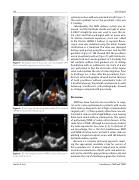
Figure 8. Fluoroscopy clip showing deployment of the inverted SAPIEN 3 valve in the mitral position.
Figure 9. TEE showing the inverted SAPIEN 3 valve in the appro- priate mitral position.
optimal position without paravalvular leak (Figure 7). The mean gradient across the prosthetic valve was 5.7 mmHg.
Subsequently, the TAVR delivery system was re- moved, and the Certitude sheath was kept in place. A 0.035” straight-tip wire was used to cross the mi- tral valve and then exchanged with an Inoue wire. To achieve maximum expansion, 4 mL was added to the 29-mm SAPIEN 3 balloon. A coplanar uoro- scopic view was obtained using the mitral annular calci cation as a landmark. The valve was deployed during rapid pacing using uoroscopic and live TEE guidance (Figure 8). TEE showed that the prosthesis was in an optimal position (Figure 9), with trivial para- valvular leak and a mean gradient of 3.5 mmHg. The left ventricle out ow tract gradient was 12 mmHg. Postdilation with an additional 2 mL (total of 6 mL) was performed to air the atrial side of the Sapien valve and minimize the risk of valve migration. Prior to discharge (i.e., 5 days after the procedure), trans- thoracic echocardiography showed normal function of both prostheses without paravalvular leaks. At 2-month follow-up, the patient continued to do well. Follow-up transthoracic echocardiography showed no changes compared with prior study.
Discussion
TAVR has been found to be non-inferior to surgi- cal aortic valve replacement in patients with severe atrial stenosis deemed to be at high or intermediate surgical risk [1, 2]. These patients often have concom- itant mitral stenosis with a high Wilkins score, barring them from mitral balloon valvuloplasty. The option of performing TMVR of native mitral stenosis at the same time as TAVR, although not previously studied, has been reported in two cases [5, 6]. To the best of our knowledge, this is the rst simultaneous TAVR and TMVR of native aortic and mitral valves stenoses utilizing a single transapical access with the Edwards Certitude delivery system.
Because is a complex and novel approach, select- ing the appropriate candidate is key for success of this procedure. It is of utmost importance to obtain accurate measurements of both aortic and mitral an- nuli and to select the appropriate prosthesis size and minimize the risk of interference given the anatomi-
Tandar A. et al.
Simultaneous TAVR and TMVR
