
Page 19 - Journal of Structural Heart Disease Volume 3, Issue 5
P. 19
145
Case Report
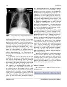
Figure 3. Post-procedure chest radiograph depicting relation- ship of vascular plug to the mechanical mitral valve ring.
advancing catheters and/or devices [2]. Creating a veno-arterial loop via trans-septal puncture, as was performed in our patient, allows for stable wire po- sitioning and permits an anterograde or retrograde approach to device closure. Although there is po- tential for hemodynamic instability with a veno- arterial loop due to the wire stenting open the aortic valve, our patient tolerated the approach well. With a pre-shaped wire loop minimizing tension across the left ventricular out ow track, it was possible to avoid hypotension related to aortic valve distortion by the wire.
An alternative approach for mitral PVL closure would be to advance the sheath directly to the left ventricle through the PVL without creating a veno-arterial loop; however, the trade-o would be attempting to advance a comparatively large sheath into a small PVL with a short length of wire of lim- ited stability in the left ventricle. We believe that in an infant with very short left ventricular length, it would be possible but unlikely that one could maintain adequate wire position and successfully advance the 4-F sheath into position through the small, tight leak. There would possibly be increased risk of wire perforation of the left ventricular apex given the small dimensions and limited room for
wire mobilization. Conversely, this approach may be more amenable with a more sizeable PVL in a larger patient. A trans-apical approach is also feasible, with the bene t of allowing direct entry of the mitral PVL regardless of its location; this can be performed per- cutaneously or open [2]. Disadvantages to a percu- taneous apical approach are that it is more invasive and requires additional pre-procedure planning and imaging (i.e., ultrasound, echocardiography, and/ or computed tomography) and that there is limited availability of dedicated apical closure devices do- mestically or internationally [5-7]. The open approach has additional limitations, such as an increased risk of apical tear, bleeding, myocardial damage, arrhyth- mia, coronary damage, and infection.
In terms of device selection, the size of the patient must be considered for obvious technical reasons. The Amplatzer ventricular septal defect device has been used in larger patients; however, due to our pa- tient’s size and risk of iatrogenic mitral valve or pul- monary vein obstruction, we utilized the Amplatzer Vascular Plug II. This proved to be a practical device for closure of a PVL in the United States, although other candidate devices are presently available do- mestically (i.e., Amplatzer Duct occluder, Amplatzer septal occlude, vascular coils) and internationally (i.e., Amplatzer Vascular Plug III and 4) [2, 8]. Newer, dedicated PVL closure devices are also on the hori- zon, such as the Occlutech PVL device, but due to their size, these would be better suited for larger de- fects in larger patients.
Successful transcatheter PVL closure has previously been described in children, adolescents, and adults. However, our report demonstrates that successful closure of a PVL in an infant under 12 months old is technically feasible, can result in clinical improve- ment of sequelae attributed to PVL, and may obviate the need for repeat surgical intervention.
Con ict of Interest
The authors have no con ict of interest relevant to this publication.
Comment on this Article or Ask a Question
Nwankwo, U. et al.
Closure of Mitral Paravalvular Leak in an Infant
