
Page 18 - Journal of Structural Heart Disease Volume 3, Issue 5
P. 18
Case Report 144
AB
C
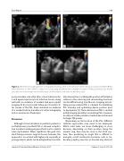
Figure 2. (Panel A) Transesophageal echocardiography showing a small-to-moderate prosthetic mitral paravalvular leak (Panel B) The defect measures 2.7 mm. (Panel C ) Status post vascular plug placement with complete resoulution of the paravalvular leak; the left atrial disk does not protrude into the left atrium. LA, left atrium; LV, left ventricle; MV, mitral valve.
post-procedure, and other than a short admission for a viral upper respiratory tract infection, he was doing well with no evidence of residual leak upon repeat imaging. At his most recent follow-up 27 months af- ter closure of the PVL, there remained no evidence of a residual leak by transthoracic echocardiography and no recurrence of hemolysis.
Discussion
Although its true incidence in pediatric patients is not fully known, prosthetic PVL is a known complica- tion in patients undergoing prosthetic aortic or mitral valve replacement. When signi cant, the gold stan- dard therapy remains surgical closure; however, this approach is associated with higher risk, especially in young patients, which can be mitigated by transcath-
eter intervention. Localizing the position of the leak in relation to the valve ring and surrounding structures can be di cult using transthoracic imaging, but uti- lizing peri-procedural TEE is valuable for identifying PVL anatomy and optimizing device position prior to deployment [4]. Three-dimensional TEE is another useful imaging modality in visualizing PVLs; however, its utility in smaller patients is limited due to the need for larger TEE probes.
Depending on the location of the PVL, di erent catheter approaches may need to be employed. Mitral valve leaks are more challenging to close because, depending on their position along the annular ring, they may be close to the atrial sep- tum, thus producing an angle that is di cult to navigate, or left ventricular structures such as tra- beculae, papillary muscles, or chordae may obstruct
Journal of Structural Heart Disease, October 2017
Volume 3, Issue 5:141-146
