
Page 99 - Journal of Structural Heart Disease Volume 4, Issue 4
P. 99
201
Meeting Abstracts
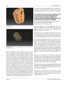
159. Figure 1. Gross Pathological Specimen of Dilated Cardio- myopathy.
159. Figure 2. Melody Transcatheter Pulmonary Valve (Medtron- ic Corportation).
3D scanning (3DS) is a novel application where the res- olution of imaging is limited only by graphical and pro- cessor capability. 3DS has potential to delineate, with previously unforeseen detail, highly complex anatomic structures. We review cases using pathologic specimens where use of 3DS enabled virtual 3D reconstruction with resolutions exceeding current clinically-available CT or MRI images. 3DS in the future can be used to scan pathologi- cal specimens to better understand the morphology and anatomy of congenital heart defects. Moreover, individual devices, pathological specimens, and animal specimens both with and without devices implanted can be scanned with the aim of designing novel devices for minimally inva- sive catheter-based interventions. Finally, not only can 3D printed models derived from imaging studies be scanned and assessed for accuracy, but di erent imaging modalities can be compared to see which produces the most accurate models. These 3D models and scans also have the second- ary bene t of educating cardiology and surgical fellows.
In conclusion, 3D scanning and printing is a valuable sup- plement to traditional methods of procedural planning and has the capacity to o er novel treatments to patients.
160. THE FIRST CASE OF SUCCESSFUL EMBOLIZATION BY A VASCULAR PLUG FOR A PARAVALVAR LEAK AFTER TRICUSPID VALVE REPLACEMENT IN A PATIENT AFTER EXTRACARDIAC FONTAN OPERATION FOR HYPOPLASTIC LEFT HEART SYNDROME
Satoshi Yasukochi, Hidehiko Hara, Kiyohiro Takigiku, Kouta Takei, Takeshi Hashimoto, Masaru Okamura
Nagano Children's Hospital, Nagano, Japan
Paravalvar leak (PVL) is a possible complication after pros- thetic valve implantation in adult which may cause sig- ni cant symptoms of congestive heart failure(CHF) and hemolysis, but very rare in child.
We report here a 14-year-old boy as the rst successful case of percutaneous closure of PVL in tricuspid prosthetic valve by a vascular plug, 11 years after extracardiac Fontan pro- cedure (eTCPC) for hypoplastic left heart syndrome(HLHS).
He had been su ering from CHF with chest pain due to PVL after TVR ( ATS Φ24mm), which underwent at 13 years old for severe progressive tricuspid regurgitation(TR) after eTCPC , whose postoperative course had a poor recov- ery. Transesophageal echocardiography (TEE) demon- strated a paravalvar defect in 3-5mm size along at 1-2 o’clock position of prosthetic TV and severe regurgitation. Hemodynamic measurements by catheterization showed elevated mean pulmonary wedge pressure (PAWP) as 12mmHg and ejection fraction of right ventricle(RV) was 32%.
Under general anesthesia, 4F customized catheter via left femoral artery was inserted retrogradely through RV into left atrium (LA) with 0.035” Terumo guide wire(GW). With TEE guide, a guide catheter in LA was changed to 5F Destination catheter with Safari GW, then Safari GW was exchanged by 0.014” Cruise GW as a leading GW.
The PLV was successfully closed by AVP-II(Φ=10mm), after failing the rst attempt by AVP-II (Φ8mm) which resulted in migration and retrieved. After closing PVL, mean LA pres- sure was down to 8mmHg.
Percutaneous closure of PVL by a vascular plug is a pow- erful alternative treatment of surgery even in a child with multiple surgery for complex heart disease like our case who limited the vascular access.
Hijazi, Z
21st Annual PICS/AICS Meeting
