
Page 90 - Journal of Structural Heart Disease Volume 4, Issue 4
P. 90
Meeting Abstracts
192
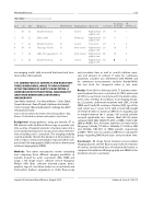
144. Table 1. Valve tricuspid valve-in-valve.
Case
Sex
Age
Diagnosis
FC NYHA pre
Regurgitation
Approach
Size Valve
Hospitalar discharge
FC
NYHA post
1 M32
2 M22
3 F42
4 M16
5 F34
6 F46
Ebstein Anomaly II
Endocarditis II
Ebstein Anomaly III Tetralogy of Fallot VI
VSD +TV regurgitation III Ebstein Anomaly III
Severe Moderate
Severe Mod-Severe
Mod-Severe Severe
Righ internal 26 19 jugular vein
Righ internal 26 11 jugular vein
Transatrial 28 7
Righ internal 28 18 jugular vein
Righ internal 30 10 jugular vein
Righ internal 30 4 jugular vein
I
I
II II
II II
encouraging results with increased functional and struc- tural cardiac improvement.
145. AIMING FOR LESS CONTRAST AND RADIATION: THREE-DIMENSIONAL IMAGE FUSION GUIDANCE
IN THE TREATMENT OF AORTIC COARCTATION: A COMPARISON WITH TRADITIONAL ANGIOGRAPHY AND THREE-DIMENSIONAL ROTATIONAL ANGIOGRAPHY.
Juan Pablo Sandoval1, Jose Garcia Montes1, Carlos Zabal1, Tomasz Moszura2, Pawel Dryzek2, Guillermo Aristizabal1, Carlos Sisniega1, Maciej Lukaszewski2, Jadwiga Ana Moll2, Sebastian Goreczny2
1Ignacio Chavez National Institute of Cardiology, Mexico City, Mexico. 2Polish Mother's Memorial Hospital, Lodz, Poland
Background: Image-guidance using pre-existent CT or MR datasets with traditional uoroscopy to provide real- time overlay of targeted anatomic structures represents a novel and promising tool in vascular and cardiac interven- tions including aortic coarctation. This imaging modality could potentially shorten the duration of interventions as well as reduce contrast and radiation exposure when com- pared with 2D angiography (2DA) and three-dimensional rotational angiography (3DRA).
Methods: Two-centre retrospective review evaluating and comparing three di erent imaging modalities in patients treated for aortic coarctation: 2DA, 3DRA and using a 3D image fusion software (Vessel Navigator, Philips) (VN). Data collected included patient demo- graphics, type of coarctation (native vs recoarctation) and intervention (balloon angioplasty or stent), uoroscopy
and procedure time as well as overall radiation expo- sure and amount of contrast. P value for continuous parametric variables was determined with ANOVA and for continuous non-parametric variables Kruskal-Wallis test was used. For categorical values χ2 was used.
Results: From 2015 to February 2018, 77 patients under- went treatment for aortic coarctation; 54 (70%) native and 23 (30%) recoarctation. Seventy-two (93%) patients under- went aortic stenting. According to each imaging modal- ity, 22 patients underwent treatment with 2DA, 24 with 3DRA and 31with VN assistance. Median (IQR) age of the total cohort was 11 years (6-15) with a mean (SD) weight of 39.9(23.4) with no statistical di erences regarding age or weight between the 3 groups. VN assisted patients received signi cantly less contrast, 90ml (50-159) when compared with 2DA, 140ml (75-207) or 3DRA, 162ml (120- 200) (p<0.001). Also, Air kerma tended to be lower in the VN group, 136mGy (77-309) vs 303mGy (116-454) in 2DA and 233mGy (103-527) in 3DRA patients respectively, p=0.089. There were no statistical di erences among the groups regarding DAP, uoroscopic and procedure time.
Conclusions: 3D image fusion between pre-procedural imaging datasets and live uoroscopy results in reduction of contrast and absorbed dose of radiation (Air kerma) as compared to traditional 2D angiography and 3DRA for the treatment of aortic coarctation.
Journal of Structural Heart Disease, August 2018
Volume 4, Issue 4:114-206
