
Page 63 - Journal of Structural Heart Disease Volume 4, Issue 4
P. 63
165
Meeting Abstracts
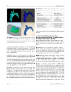
96. Figure. Generation of a 3-D printed model. DICOM data is rst segmented to de ne the anatomy of interest (a. Fontan in cyan, trachea in green.) The model is cleaned (b. Fontan in blue, trachea in green) and prepared for printing. (c). Final 3-D printed model (d. Fontan in blue, trachea white.)
cardiologists provided descriptions of the 3-D printed anatomy in addition to a subjective rating (1-5) on quality and accuracy compared to the digital models. Data were compared using Mann-Whitney U test, χ2 and κ for agree- ment, as appropriate.
Results: RA data from 13 catheterizations were 3-D printed. Patient characteristics, details of image acquisition and 3-D printing are shown in the Table. All models were of vascular structures (aorta, pulmonary arteries, Glenn/Fontan con- duits, coronary artery stula). The independent reviewers correctly described 70 and 85% of the models (p=0.077) and rated the quality and accuracy of the models high with good agreement (5 vs. 5, p=NS; κ=0.458).
Discussion: This proof of concept study has shown that DICOM data from standard RA can be successfully con- verted into 3-D printed models with good accuracy for de ning anatomy. The cost of printing the models was negligible, but the time to print is still too long to allow for real-time use of the models. As the speed of 3-D print- ing technology increases, a novel future application of this technique could allow for printing of patient-speci c
96. Table. Demographics, details of image acquisition and 3-D printing. Data are presented as median (interquartile range) or n (%).
stents and devices in the catheterization lab based on RA datasets.
97. TRANSCATHETER DEVICE CLOSURE OF AORTOPULMONARY WINDOW. IS IT A REASONABLE ALTERNATIVE TO SURGICAL REPAIR?
Keneth Magaña, Aldo Campos, José Garcia Montes, Carlos Zabal, Jorge Cervantes, Juan Calderon, Juan Pablo Sandoval Ignacio Chavez National Institute of Cardiology, Mexico City, Mexico
Background: An aortopulmonary window (APW) is a defect caused by incomplete separation between the aortic and pulmonary wall during early embryogenesis. Classical treatment is surgery with only few reported cases undergoing transcatheter device closure (TDC).
Methods: Single center, retrospective analysis including children with APW who underwent TDC as an alternative to surgery between 2004 and 2017. Patients were compared to those undergoing surgery during the same period. Clinical, interventional and surgical aspects were reviewed.
Results: Six children underwent TDC of APW. Median age was 4 years (1month-15years) and weight was 30 kg (3-45). Defects considered suitable for device closure were all type I defects according to the Mori et al. classi cation. Median diameter of the defect was 6.7 mm (2.31-14.9). Median systolic pressure of the pulmonary artery (sPAP) prior to closure was 50 mmHg (25-80); PVRi was 3.7UW (2.1-4.5); Qp:Qs of 3:1(1.5-4.56). APW’s were closed using an Amplatzer Duct Occluder (n=3); ADO II (n=1), Cera PDA occluder (n=1), and an Amplatzer Septal Occluder (n=1). Device embolization occurred in two children (33%) within 24 hours post implantation. In both cases, devices
Hijazi, Z
21st Annual PICS/AICS Meeting
