
Page 21 - Journal of Structural Heart Disease Volume 4, Issue 4
P. 21
123
Meeting Abstracts
2 Department of Cardiac Surgery, Hiroshima City Hospital, Hiroshima, Japan
A baby boy was born via emergent cesarean section at 26 weeks of gestation, weighing 438 g. Echocardiography revealed a large PDA and valvular aortic stenosis (AS) of 1.9m/s, requiring PDA ligation at 5 days of age. He rapidly developed systemic edema and bradycardia with motion when he was 111 days old and 890 g. Echocardiography revealed an AS of 5.1 m/s, LVEDD of 219%, LVEF of 54%, and no patency of the foramen ovale. The emergent interven- tion was required. The ultrasonography revealed that the RCCA (2.7 mm) could accommodate the sheath, although the femoral artery was too small (1.6 mm). The patency of the anterior communicating artery was proved which maintained blood perfusion to the brain even the RCCA might be obstructed by the sheath. The length needed for the sheath was measured by determining the distance between the point of insertion and AoV –then the length between AoV and LV apex (must be longer than half the length of balloon plus balloon shoulder length). Silkworm- gut was tied 20 mm from the tip of the sheath to ensure the sheath was not inserted too far. In a hybrid operating room, the surgeon made a semicircle incision on the RCCA and a 4-Fr sheath was inserted. We avoided the pull-back gradient measurement and left ventriculography before BAVP due to critical condition. An ascending aortography revealed the diameter of AoV, and ori ce of AoV were rec- ognized using a negative jet of contrast medium. A 0.014’ guidewire was inserted into LV and BAVP was performed immediately using TMP-Ped/4 mm without raid pacing. The Pull-back pressure gradient between LV and Ao after BAVP was 22 mmHg after BAVP and cardiac function and edema rapidly improved. His prognosis was very good fol- lowing the procedure and a second BAVP was performed by antegrade approach through the femoral artery when he was 11 months old and 3.2 kg. And the angiography revealed the patency of RCCA. When performing complex procedures, simulation should be used to evaluate the pro- cedure, determine possible risks, and create strategies to ensure the best possible outcome.
21. PERCUTANEOUS CLOSURE OF PERIMEMBRANOUS VSD IN INFANTS AND CHILDREN USING AMPLATZER DUCT OCCLUDER I ; SINGLE CENTER EXPERIENCE
Hala Moustafa, Hala Hamza, Amal El Sisi, Sonia El Seidi, Aya Fatouh, Rodina Sobhy, Doaa Abd El Aziz, Wael Attia
Cairo University, Cairo, Egypt
Background/aim: Amplatzer duct occluder I (ADO I) devices appear to be an attractive option in perimembranous
(pmVSD) type. In developing countries, the lack of early available and a ordable surgery, and the relatively high cost of currently devicesdesigned for VSD closure create additional problems. We report on the immediate and mid-term follow-up results of using ADOI devices to close pmVSDs in a consecutive series of young patients.
Patients & Methods: Retrospective case note review of all children referred for transcatheter closure of pm VSD using the ADO I device.
Clinical inclusion criteria: at least 3 of the following had to be present: Overt heart failure, Failure to thrive, Recurrent respiratory infections, C/T ratio ≥0.55, LA/AO > 1.5, LVEDD z-score indexed to body surface area of ≥2, QP/QS >1.5 at cardiac catheterization, 8History of IE related to the VSD.
Morphologic inclusion criterion: Isolated pm VSD, up to 10 mm minimum diameter by TTE.TEE: to describe the anatomical position of the VSD, the distance to import- ant structures such as the aortic valve, the diameter of the defect at the LV and RV sides, the shape of the defect and the presence of tricuspid tissue from the septal lea et try- ing to estimate the needed device size
Conclusion: the transcatheter closure of pm VSD with Amplatzer ductal occluder I was successful in 95% without
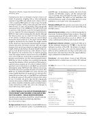
21. Table: Characteristic of the 28 studied patients who underwent VSD closure.
Variables
Median (range)
Age (yrs) Wt (kg
VSD diameter angio(mm) Fluoro time (min)
Device diameter/length
6 ×4 8 ×6 10 × 8
Procedure results
Variables
Success rate
Unstable device Embolized to RV & percutaneous retrieval
Immediate closure Closure at 3months
4 (13 months–12Y 15 (6.5–51)
5.2 (3.75–9) 55 (34.5–99) N(%)
5 (17.9%)
8 (28.6%) 13 (46.4%)
N(%)
24/28 (85.7%)
3/28 (10.7%)
1/28 (3.5%)
22/24 (91.7%) 23/24 (95%)
Hijazi, Z
21st Annual PICS/AICS Meeting
