
Page 32 - Journal of Structural Heart Disease Volume 3, Issue 6
P. 32
Case Report 188
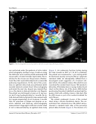
Figure 1. TEE showing bidirectional shunt before 20-mm Occluder Septal Helex placement.
was performed under the guidance of intra-cardiac echocardiography and uoroscopy. A balloon sizing the defect was used, and the patient underwent ASD closure with a 25-mm Occluder Septal Helex. The in- dication for this closure was the presence of bidirec- tional ow that would ultimately lead to irreversible pulmonary hypertension. At the end of the proce- dure, an agitated saline study was performed, which revealed minimal residual shunt. Echocardiography performed the next day showed very minimal ow across the device as demonstrated by color Doppler. The patient was discharged to home on 81 mg aspi- rin and 75 mg clopidogrel daily for 3 months. He ini- tially demonstrated symptomatic improvement with less oxygen requirement at rest. However, 3 months later, his symptoms of fatigue and dyspnea on ex- ertion returned, and follow-up echocardiography demonstrated a large right-to-left intra-cardiac shunt at rest as demonstrated by an agitated saline study
(Figure 2). His pulmonary function testing during this time showed an FEV1 of 1.78 (47% predicted). The patient was monitored for 1 year, during which his functional capacity and cystic brosis symptoms remained stable. He subsequently su ered from a neurological event suggestive of a transient isch- emic attack of an embolic nature, after which he was considered for residual shunt closure. TEE revealed a deformity of the Helex device causing a bidirectional shunt via the inferior rim of the device. With further questioning, the patient admitted to using a high-fre- quency chest wall oscillation device a few days after his ASD closure as part of his routine treatment for cystic brosis.
The patient underwent closure of the residual shunt using a 30-mm Cardioform device. The sec- ond device was advanced across the defect and po- sitioned to sandwich both sides of the previously placed Helex Occluder (Figure 3). The absence of a
Journal of Structural Heart Disease, December 2017
Volume 3, Issue 6:187-191
