
Page 11 - Journal of Structural Heart Disease Volume 3, Issue 6
P. 11
167 Original Research Article
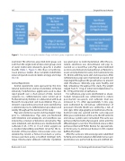
Figure 1. Flow chart showing the number of pigs and stents, group assignment, and study progression.
examined. The other two pigs (mid-term group; one each from the single stent and pre-stent groups, total of seven stents) were allowed to grow for 2 another months (Table 1, Figure 1). We chose 2 months be- cause previous studies show complete endotheliali- zation of injured vessels in rabbits and pigs at 28 days [12-14].
Animal Experiments
Animal experiments were approved by the Insti- tutional Animal Care and Use Committee at Purdue University. Yorkshire Cross piglets were used to allow rapid growth over a short period of time. Survival surgeries (i.e., catheterizations) were carried out at Purdue University facilities in collaboration with Cook Research Incorporated and Cook Medical. Pigs un- derwent a quarantine period and were examined by a veterinarian prior to catheterization procedures and weekly throughout the duration of the study.
Pigs were started on aspirin and clopidogrel 3 days prior to catheterization. Pigs were pre-medicated with tiletamine and zolazepam, and anesthesia was induced by a mixture of ketamine and xylazine. Pigs were intubated, and anesthesia was maintained with iso urane (1.25–1.75% in 1.5–2.5 L/min oxygen) via a standard rebreathing anesthetic circuit for the re- mainder of the procedures. Intravascular access was obtained by carotid or femoral cut-down or percu- taneous puncture using a modi ed Seldinger tech- nique. Intramuscular antibiotic (ceftiofur crystalline)
was given prior to stent implantation. After the pro- cedure, anesthesia was discontinued, and pigs re- covered on a raised oor pen. Pigs were medicated as necessary (meloxicam, butorphanol, or unixin) to assure uncomplicated recovery and monitored every 15–30 min until they were alert and responsive. After catheterization, pigs were maintained on aspirin and clopidogrel throughout the growth phase to prevent stent thrombosis. Clinically, pigs were healthy and fully ambulatory throughout the study. Pig weight ranged from 9–11 kg at initial stent implantation to 98–119 kg at the time of euthanasia.
For euthanasia, pigs were anesthetized as above. Sodium nitroprusside was administered to reduce post-mortem vasospasm, and iso urane level was increased to 5%. After approximately 5 min, pigs were euthanized by intravenous administration of potassium chloride. Death was veri ed by a lack of vital signs. After angiography and euthanasia, the ab- dominal cavity was opened and the aorta exposed. After gross examination of the aorta, the left ventricle and venous systems were cannulated. The aorta was perfusion- ushed with physiological saline until the e uent began to run clear. The stented area was then perfusion- xed with Prefer xative for approximate- ly 10 min prior to immersion xation in 10% neutral bu ered formalin.
After the aorta was removed, pigs were submitted for full postmortem evaluation. Detailed macroscopic examination was performed by a board-certi ed vet-
Bratincsák A. et al.
Pre-Stenting with Intentional Stent Fracture
