
Page 33 - Journal of Structural Heart Disease Volume 3, Issue 5
P. 33
159 Case Report
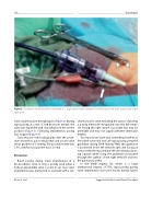
Figure 2. Pacemaker electrode (arrow) connected to a large suture needle (between arrowheads) placed trans-cutaneously in the right groin.
trans-cutaneously in the right groin (Figure 2). During rapid pacing at a rate of 160 beats per minute, the valve was expanded with slow in ation in the correct position (Figure 3). Following implantation, pacing was stopped (Figure 4).
Trans-thoracic echocardiography after the proce- dure showed no para-valvular leak and a trans-valve mean gradient of 5 mmHg. The procedure time was 1.5 h, and uoroscopy time was 12.5 min.
Discussion
Rapid pacing during valve implantation in a bio-prosthetic valve or ring is usually used when a balloon-expandable valve is used. In our case, valve implantation was performed in a patient with a me-
chanical aortic valve, excluding the option of placing a pacing electrode retrogradely into the left ventri- cle. Pacing the right atrium is possible but may be unreliable and may not supply su cient ventricular rhythm.
Two reports by Faurie et al. and Hilling-Smith et al. described successful and safe rapid pacing using the guidewire during TAVR. During TAVR, the guidewire is positioned in the left ventricle apex and has good contact with the myocardium. We describe good pac- ing capture while using the guidewire positioned through the out ow of the right ventricle and into the pulmonary artery.
In the VIVID registry [9], which is a large international registry of TTVI, rapid pacing during valve implantation was used mostly during Sapien
Eitan, A. et al.
Suggestion for Easier and Faster Procedure
