
Page 23 - Journal of Structural Heart Disease Volume 3, Issue 5
P. 23
149
Case Report
was uneven narrowing of the aortic arch between the left common carotid artery and left subclavian artery to 2.4-2.8 mm. The isthmus of the aorta immediate- ly after the ostium of the left subclavian artery had a diameter of 4.3 mm. The descending thoracic aorta had a diameter of approximately 8 mm, and the di- ameter of the thoracic aorta at the diaphragm level was 6.7 mm.
Given the high risk of open surgery for reconstruct- ing the aortic arch, a decision was made to perform endovascular intervention. However, the patient’s low weight and need for implantation of a stent that could be expanded as the body grows limited the use of access through the femoral artery. Therefore, a hy- brid approach was proposed.
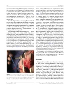
The hybrid procedure was performed in a cardio- surgical operating room using a mobile angiocardio- graphic unit OEC 9900 (General Electric Healthcare, Li- vonia, MI, USA). The patient underwent posterolateral thoracotomy at the 6th intercostal space on the left. A descending aorta segment was isolated, on which a purse string suture was applied. Through the count- er-aperture, 3 cm below the operation eld, a 6-F Mullins delivery system (NuMed, Cornwall, ON, Can- ada) was transcutaneously inserted into the thoracic descending aorta (Figure 2). The delivery system was
Figure 2. Access site through lateral thoracotomy in the 6th in- tercostal space during the hybrid stenting procedure. The deliv- ery catheter (arrow) was advanced through the thoracic aorta through a counter-aperture 3 cm below the operation eld.
carried on the guidewire to the initial section of the descending thoracic aorta through the lateral port, at which angiography of the aortic arch was performed (Figure 3A). A prolonged narrowed segment of the arch and isthmus of the aorta with a diameter of 2.4- 3.0 mm and length of 16 mm was revealed, which was located right after the origin of the left common carotid artery and extended to the descending tho- racic aorta. There was a 58 mmHg gradient across the narrowing. A decision was made to perform stenting of the entire obstructive segment using a Valeo stent that was 7.0 mm in diameter and 18 mm in length. Using a 0.014” guidewire, the right coronary catheter (JR 3.5) was advanced into the ascending aorta. The guidewire was exchanged for an 0.035” Amplatzer super sti guidewire with a 1-cm soft tip (Boston Sci- enti c, Marlborough, MA, USA) to deliver the stent into the arch of the aorta so that it would completely cover the zone of obstruction. The correct location of the stent was monitored by injecting a contrast agent through the side arm of the delivery sheath. The stent was deployed at 10 atm. Repeat angiography demon- strated very good results with complete coverage of the narrowed segment (Figure 3B). There was no gra- dient between the ascending and descending aorta. The delivery catheter was removed from the aorta, and hemostasis was achieved. The chest tube was left in place, and the incision was closed.
There were no complications during the postoper- ative period. The child was extubated after 10 h and discharged home on postoperative day 7 in good condition and on no cardiac medications.
Discussion
Kutty et al. reported six cases of aortic arch stent- ing after initial surgical treatment of hypoplastic left heart syndrome with an average age of 5.6 months (0.5-12.9 months) and weight of 5.8 kg (2.9- 7.7 kg). Stents were implanted using the ascending aorta access approach [12] and required repeated dilatations depending on the child’s growth at an average of 17.6 months (6.3-33.6) months after operation. Schmitz et al. performed stenting of the aortic arch in five pediatric patients with good clinical results; they also used the ascending aorta access approach and stated that stent implantation
Pursanov, M. G. et al.
New Approach for Hybrid Stenting of the Aortic Arch
