
Page 16 - Journal of Structural Heart Disease Volume 3, Issue 5
P. 16
Case Report
142
subsequently underwent cardiac catheterization at 4 months of age. During catheterization, he was found to have severe mitral stenosis with a mitral valve gra- dient by planimetry (mean gradient of 13-16 mmHg), a capillary wedge pressure to left ventricular end-di- astolic pressure gradient of 16 mmHg, as well as sec- ondary pulmonary hypertension with a pulmonary vascular resistance of 7.1 indexed Wood units that was poorly responsive to vasodilator therapy. Balloon valvuloplasty of the mitral valve was performed to alleviate left atrial hypertension, but there was negli- gible improvement in the valve gradient, and the pa- tient developed severe mitral insu ciency post-val- vuloplasty. He had a prolonged post-catheterization hospitalization with di culty weaning from mechan- ical ventilation as well as recurrent respiratory distress and feeding intolerance recalcitrant to medical man- agement. As his anatomy was deemed not amena- ble to surgical repair, he underwent mitral valve re- placement with a 16-mm Medtronic open-pivot AP 360 mechanical aortic valve in the supra-annular mi- tral position at 5 months of age. His post-operative course was complicated by a small PVL rst noted upon post-operative transesophageal echocardiog- raphy (TEE). In addition, he developed a moderate pericardial e usion that eventually required surgical intervention with a pericardial window. The PVL was noted by all subsequent TEE and was localized to the posterolateral ring of the valve. There was no wors- ening of the leak upon serial imaging; however, the patient was readmitted for an elevated international normalized ratio and was found to have developed persistent hemolysis with elevated plasma-free he- moglobin and lactase dehydrogenase levels, requir- ing repeated blood transfusions. Attempted medi- cal therapy with pentoxifylline failed, thus 2 months post-operatively, the patient was referred to the car- diac catheterization laboratory for percutaneous PVL closure.
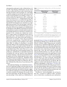
Hemodynamic ndings are summarized in Table 1. The most signi cant ndings were elevated left atrial pressure, elevated mitral valve peak gradient, and el- evated left ventricular end-diastolic pressure sugges- tive of diastolic dysfunction. Procedural TEE identi ed a small-to-moderate PVL between the mouth of the left atrial appendage and the left upper pulmonary vein measuring 1.2 mm, which was con rmed by an-
Table 1. Hemodynamic ndings before and after paravalvular leak closure.
RA 5/7/4
RV 42/3 – MPA 32/14/23 – RPA 30/11/21 – RPAW 17/16/14 – LPA 35/15/25 – LPAW 17/12/17 –
Site
Before Closure (in mm Hg)
After Closure (in mm Hg)
LA 20/18/14 LV 88/14 AAO 82/38/57 DAO 71/38/53
8/10/7
15/20/13 98/12 90/40/62 71/39/54
AAO = ascending aorta; DAO = descending aorta; LA = left atrium; LPA = left pulmonary artery; LPAW = left pulmonary artery wedge pressure; LV = left ventricle; MPA = main pulmonary artery; RA = right atrium; RPA; right pulmonary artery; RPAW = right pulmonary artery wedge pressure; RV = right ventricle.
giography (Figure 1A, Figure 2A and 2B). A trans-septal approach was utilized to access the left atrium. Using a Cobra C1 catheter, a 0.018” oppy wire was advanced from the venous sheath through the PVL and into the ascending aorta, where it was snared by a B-Braun mul- tisnare introduced from the arterial sheath. This in ef- fect created a veno-arterial loop of wire from the right femoral vein, across the atrial septum, through the leak, into the left ventricle, across the aortic valve, and into the descending aorta to the right femoral artery (Figure 1B). Using a 4-F Mullins sheath advanced trans- venously across the PVL, a 4 mm × 6 mm Amplatzer Vascular Plug II was then positioned anterogradely and was successfully deployed within the tract. Place- ment of the vascular plug was assisted by TEE. No signi cant residual leak into the left atrium was visu- alized by angiography or TEE (Figure 1C, Figure 2C). Post-intervention, both the left atrial pressure and mi- tral valve peak gradients decreased. Total uoroscopy time was 54 min, and the procedure time was 2 h and 39 min. The patient tolerated the procedure well, and there were no associated complications.
Transthoracic echocardiography on post-proce- dure day 1 showed no evidence of PVL, and there was no mitral valve or pulmonary vein obstruction.
Journal of Structural Heart Disease, October 2017
Volume 3, Issue 5:141-146
